
Basics of dermatology
2. Fundamentals of Dermatology
2.1 Structure and Function of the Skin
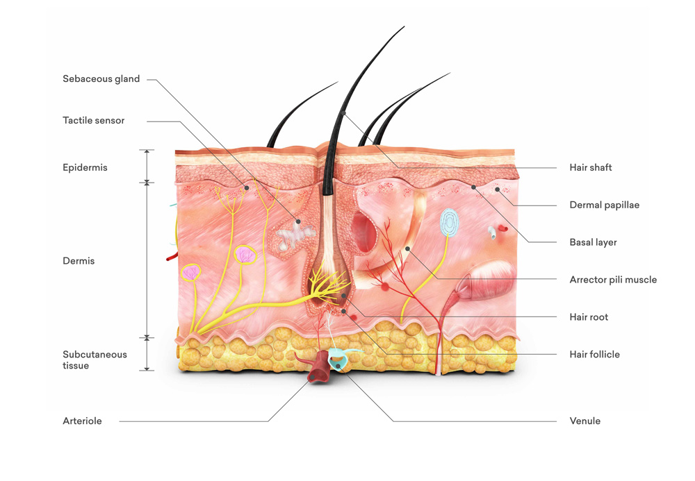
The skin (Latin: cutis; Greek: derma) is the largest and most functionally versatile organ in the human body. It covers an area of about 1.7 to 2 square meters and weighs approximately 3.5 to 10 kilograms, depending on body size and body fat percentage. The skin consists of three main layers: the epidermis (outer layer), the dermis (middle layer), and the subcutis (underlying layer).
2.1.1 Epidermis (outer layer of the skin)
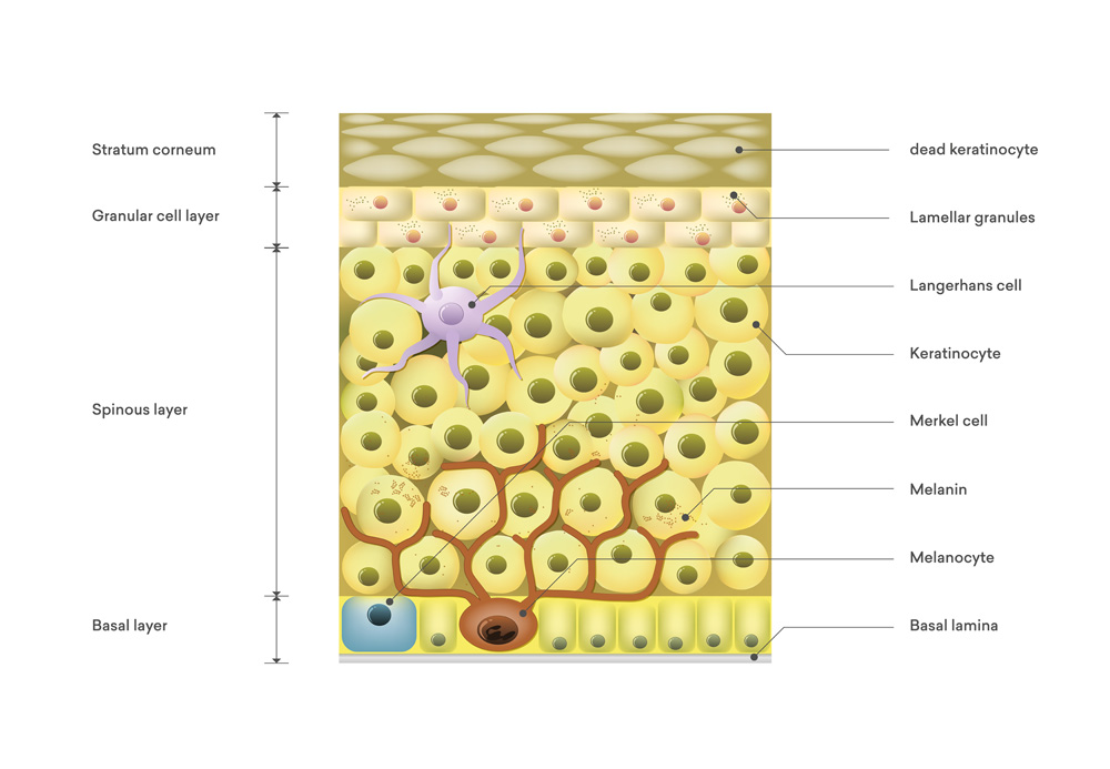
The epidermis is the outermost layer of the skin and consists mainly of keratinocytes. These cells produce keratin, a protein that gives the skin its firmness and resilience. The epidermis has no blood vessels and is nourished by the diffusion of nutrients from the underlying dermis.
- Stratum corneum (horny layer): The outermost layer, consisting of dead, keratinized cells. It protects against external factors and water loss.
- Stratum lucidum (lucid layer): A thin, clear layer found only in the skin of the palms and soles.
- Stratum granulosum (granular cell layer): Here, the cells begin to keratinize and form dense keratohyalin granules.
- Stratum spinosum (spiny cell layer): This layer contains cells with spiny projections that are connected by desmosomes and provide stability to the skin.
- Stratum basale (basal cell layer): The deepest layer of the epidermis, consisting of dividing cells (stem cells) that are responsible for the regeneration of the epidermis.
2.1.2 Dermis (skin)
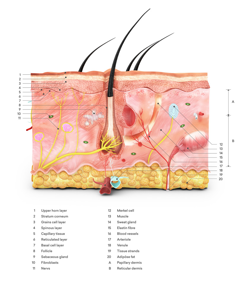
The dermis lies beneath the epidermis and consists mainly of connective tissue. It contains blood vessels, nerves, hair follicles, sebaceous glands, and sweat glands. The dermis is divided into two layers:
- Stratum papillare (papillary layer): This superficial layer contains fine, elastic fibers and forms papillae that protrude into the epidermis, providing nourishment to the epidermis and ensuring its mechanical stability.
- Stratum reticulare (reticular layer): The deeper layer of the dermis, which consists of dense, mesh-like collagen fibers and gives the skin its firmness and elasticity.
2.1.3 Subcutis (Subcutaneous Tissue)
The subcutis consists of loose connective tissue and fat cells (adipocytes). It serves as a cushion, an energy store, and an insulating layer. The subcutis connects the skin to the underlying structures, such as muscles and bones.
2.2 Functions of the Skin
The skin performs numerous vital functions:
- Protective function (Protectio): The skin protects the body from mechanical stress, chemical substances, microorganisms, and UV radiation.
- Temperature Regulation (Thermoregulation): Through sweating and the regulation of blood flow, the skin helps regulate body temperature.
- Sensory function (sensitivity): The skin contains numerous receptors that detect stimuli such as pressure, touch, temperature, and pain.
- Metabolic function (metabolism): The skin is involved in the synthesis of vitamin D and plays a role in fat metabolism.
- Immunological function: The skin contains Langerhans cells, which are part of the immune system and help defend against pathogens.
- Communication: The skin often reflects the body's emotional and physical state (e.g., blushing, turning pale).
2.3 The Layers of the Skin in Detail
2.3.1 Epidermis (Outer Layer of the Skin)
The epidermis is a multilayered, keratinizing squamous epithelium. The cells undergo a continuous renewal process that lasts approximately 28 to 35 days.
- Stratum corneum: It consists of 15–20 layers of dead, keratinized cells that form a protective barrier against environmental factors and water loss.
- Stratum lucidum: This layer is found only in the palms of the hands and the soles of the feet, and it gives these areas their unique strength and resilience.
- Stratum granulosum: This is where the keratinization process begins; the cells contain granules of keratohyalin.
- Spiny layer: The cells are connected by desmosomes, which provide the skin with additional mechanical stability.
- Basal stratum: This layer contains dividing basal cells, which are responsible for the regeneration of the epidermis.
2.3.2 Dermis (skin layer)
The dermis is a thick layer of connective tissue that supports and nourishes the epidermis. It consists of two main layers:
- Papillary layer: This superficial layer contains fine elastic fibers and papillae that interlock the dermis with the epidermis and ensure a good blood supply.
- Reticular layer: This deeper layer contains thicker collagen fibers and elastic fibers, which give the skin firmness and elasticity. Most of the glands and hair follicles are also located here.
2.3.3 Subcutis (Subcutaneous Tissue)
The subcutis consists mainly of adipose tissue and loose connective tissue. It serves as an energy store, provides cushioning against mechanical stress, and acts as insulation against the cold. The subcutis connects the skin to deeper structures such as muscles and bones.
2.4 The Basal Membrane
2.4.1 Function of the Basal Membrane
- Protection: The basement membrane serves as a protective layer between the epidermis and the dermis. It prevents foreign substances, including pigments, from penetrating uncontrollably into the deeper layers of the skin, where they could cause more serious damage.
- Pigmentation: In permanent makeup, pigments are deposited into the epidermis and the uppermost layer of the dermis. This is done using special needles that precisely inject the pigments into the correct layer of skin, where they can remain for a long time.
- Skin Regeneration: The basement membrane plays an important role in skin regeneration. It supports the formation of new skin cells and the continuous renewal of the epidermis. This regeneration is crucial for ensuring that permanent makeup lasts a long time and that the skin remains healthy.
2.4.2 Meaning for permanent makeup
- Shelf life: The pigments, which are deposited in the epidermis and the outermost layer of the dermis, remain there for several years and gradually fade. This results in a long-lasting effect that, unlike traditional tattoos, penetrates less deeply into the skin and therefore poses fewer health risks.
- Technology: The correct technique for applying the pigments is crucial. The needles must work with precision to place the pigments in the optimal layer of skin. This requires expertise and experience to ensure that the pigments are distributed evenly and to achieve the desired aesthetic result.
- Risks: Improper procedures can lead to complications such as inflammation, infection, or scarring. It is therefore of the utmost importance that permanent makeup be performed by qualified professionals who are thoroughly familiar with the techniques and potential risks and who adhere to appropriate hygiene standards.
Summary:
The basement membrane is a critical structure that separates the epidermis from the dermis and plays an essential role in skin regeneration. For permanent makeup, the proper placement of pigments in the epidermis and the uppermost layer of the dermis is crucial for the longevity and quality of the results. Proper treatment and technique are essential to ensure long-term durability and minimize risks.