Dermatology

Dermatology
The skin (gr. derma; lat. cutis) is the most functionally versatile organ of the human organism. As an envelope organ, it protects against solar radiation, heat loss and environmental influences. The skin has a wide range of adaptation mechanisms and performs self-regulation tasks as well as important metabolic and immunological functions. It also serves to absorb sensory stimuli such as vibration, temperature and pain.
The skin is a protective barrier against external influences and shields the inside of the body from the environment. However, even in healthy skin, dissolved substances can penetrate along the hair follicles into the deeper layers of the skin. This can be used to our own advantage, for example in the treatment of many skin diseases. However, this mechanism is also the source of a wide range of damage.
The skin also serves as a means of representation and communication. It communicates emotions to the outside world by blushing, paling and "ruffling the hair". It is also said that the skin is "the mirror of the soul". The skin also sends odor messages via scents (pheromones).
Structure of the skin
An adult human has a total skin area of around 1.73 m² with a weight of around ten to 14 kg. The thickness varies between 1.5 and 4 mm depending on the area of skin.
The outer skin is divided into three main layers: Epidermis (epidermis), Dermis (dermis, lat. Corium) and Subcutis (subcutis). The epidermis and dermis together form the cutis.
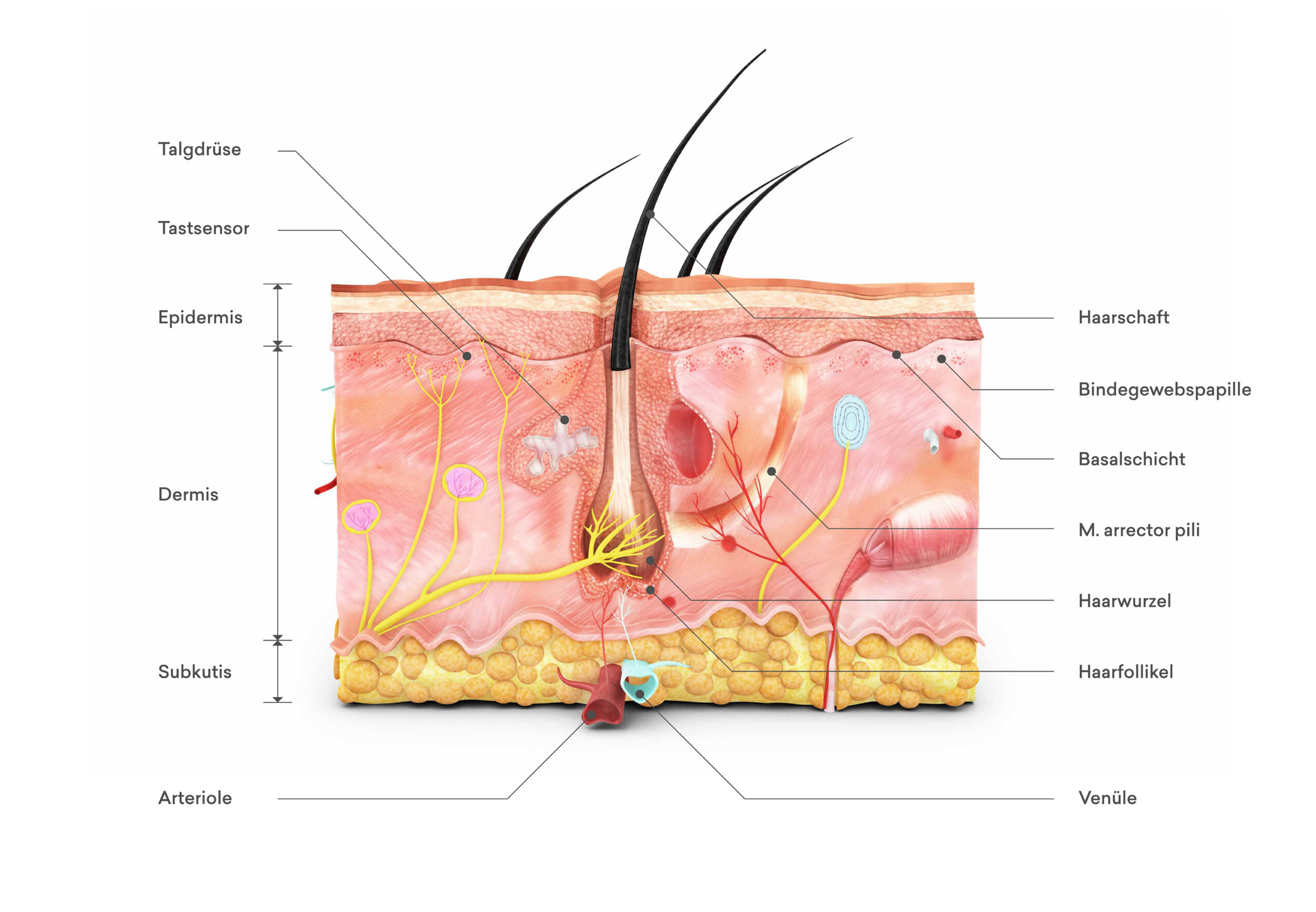
Epidermis (epidermis)
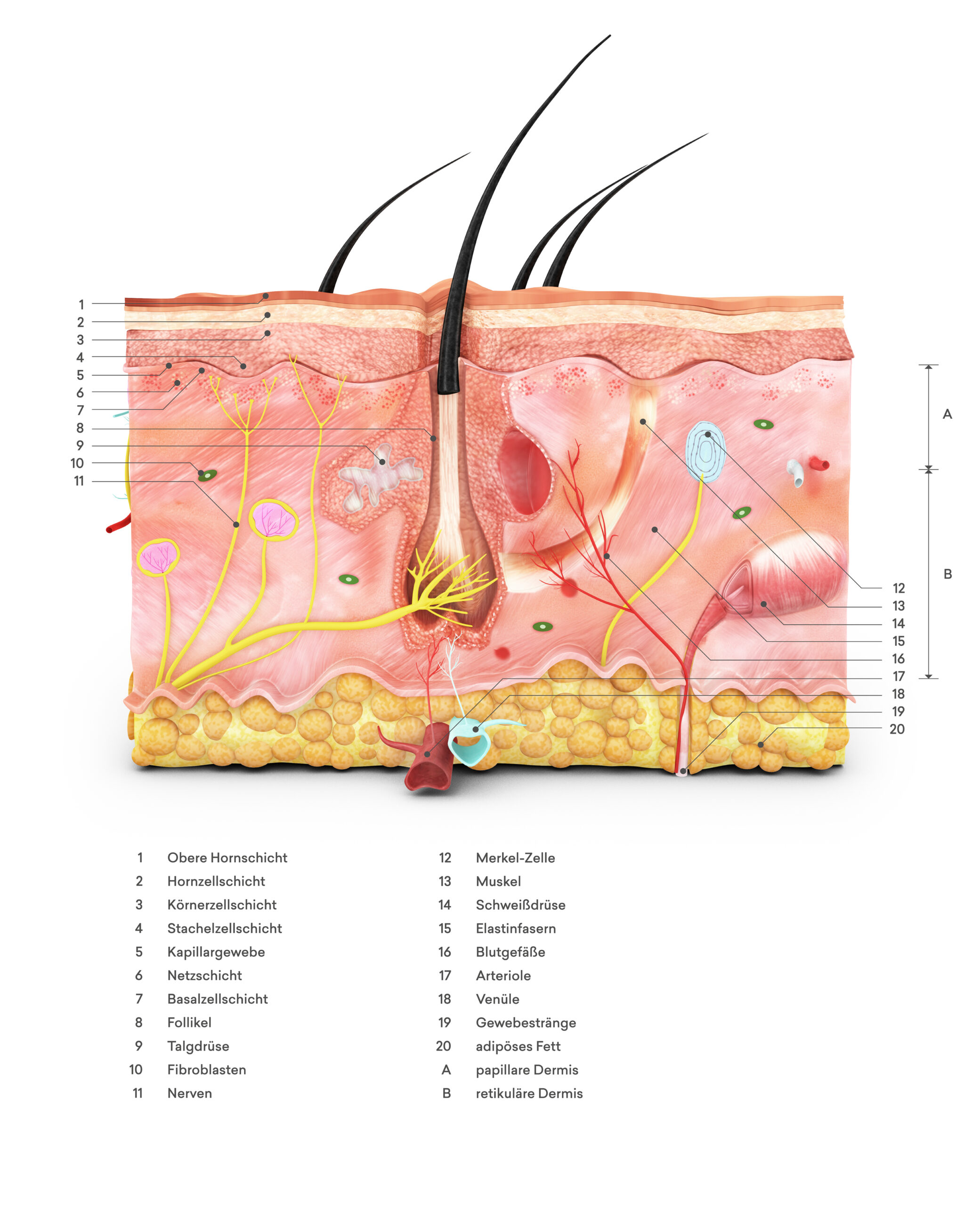
The epidermis belongs to the epithelial tissues. It is a multi-layered, keratinizing squamous epithelium. Depending on the skin area and condition, it consists of 15 to 20 closely connected cell layers. The thickness of the epidermis is between 0.03 and 0.05 millimetres, depending on the body part and function.
From the outside to the inside, the structure of the epidermis is divided into the following layers:
- horny layer (Stratum corneum)
- Glossy layer (Stratum lucidum) - is only present on the inguinal skin of the palms of the hands and feet
- Grain cell layer (Stratum granulosum)
- Spiny cell layer (Stratum spinosum)
- Basal cell layer (Stratum basale)
The spiny cell layer and the basal cell layer together form the germinal layer (Stratum germinativum).
The basement membrane is part of the epidermis and separates it from the dermis. This layer is responsible for the reproduction of skin cells. As soon as new cells have formed, they replace the overlying skin cells, which are shed. In this way, the epidermis is renewed within a cycle of 28 to 35 days. This continuous cell renewal makes it possible for the skin to heal completely after injuries.
The natural pigmentation of the skin also takes place in the basement membrane. However, pigments can also be introduced here on a long-term basis. Due to the constant skin renewal, the skin can fully regenerate after a properly performed pigmentation.

Dermis (dermis, corium)
The dermis consists mainly of connective tissue fibers (collagen and elastin fibers) and serves to nourish and anchor the epidermis. It is divided into the Stratum papillare (papillary layer, cone layer, papillary body) and into the Stratum reticulare (mesh layer).
The dermis is elastic, it can stretch and relax. The thickness of the dermis is 1 to 4 mm, depending on the part of the body. All glands, nerves and vessels are located in the dermis. The skin appendages also originate from the dermis. These include hair with its sebaceous glands and the hair follicle muscle (Arrector pili muscle), nails and all other sebaceous and sweat glands. The lower dermis contains the smooth muscles, which are important for temperature regulation, and the blood vessels.
Subcutis (under the skin)
The subcutis forms the base for the overlying skin layers and contains blood vessels and nerves as well as subcutaneous fat and loose connective tissue. This layer of skin is not relevant for pigmentation: If color were to stray into this layer of skin, it would disappear into the fatty tissue.
Wound healing
If the skin is injured, the body endeavors to restore the lost protection as quickly as possible. This rapid wound healing is ensured by cells, some of which are directly in the skin and some of which are transported to the injured skin area with the blood.
What happens in the skin during pigmentation?
When pigmentation is carried out properly, skin cells are displaced, stitch channels are opened and some are cut superficially. You are at most in the upper area of the dermis.
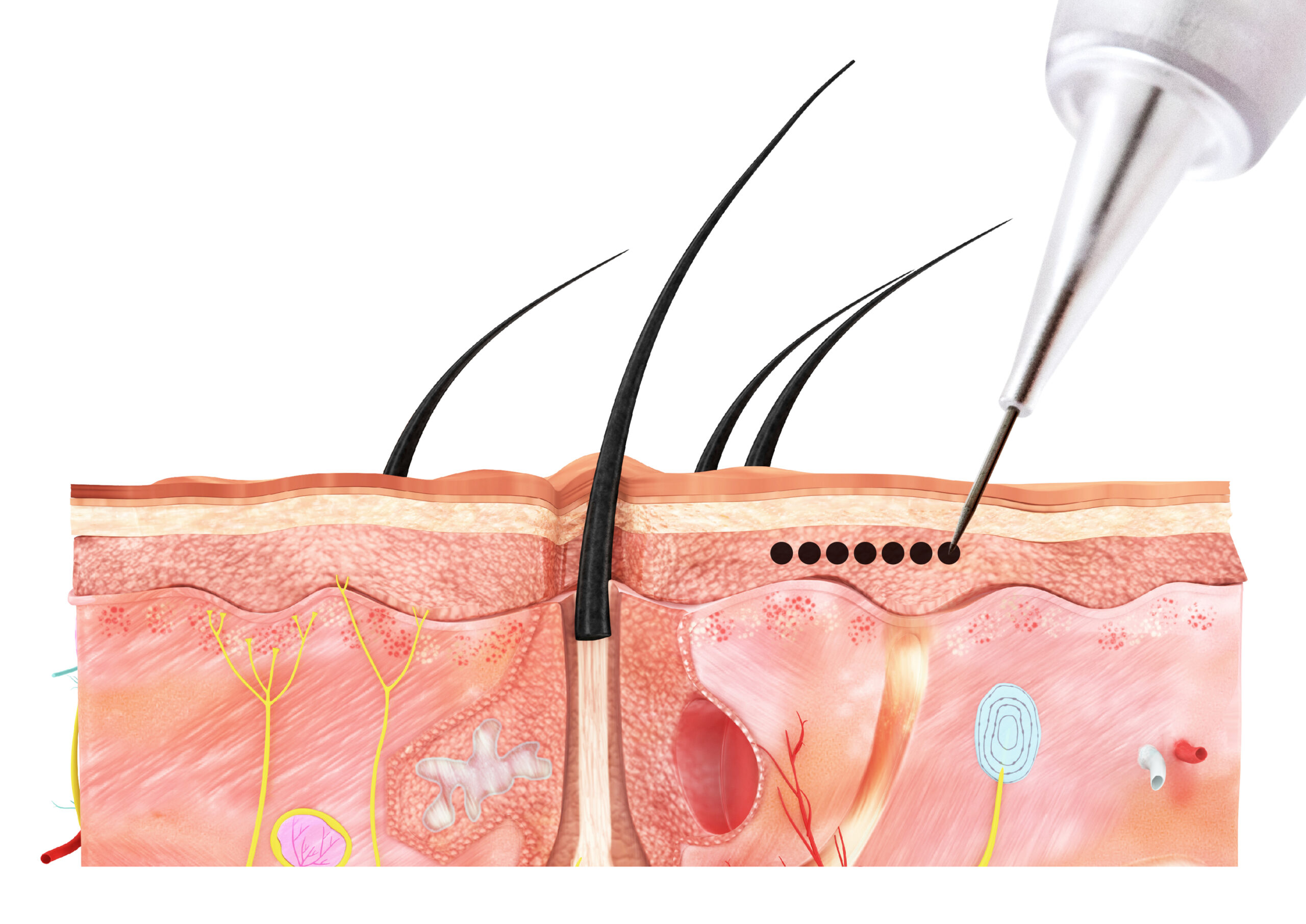
These superficial wounds, in which the epidermis and at most the uppermost layer of the dermis are injured, heal regeneratively. If the basement membrane remains intact, complete regeneration can be assumed: This means that the skin regenerates without scars.
A prerequisite for regenerative wound healing:
- The wound is kept clean and not contaminated with germs or foreign bodies.
- The instructions for aftercare are properly followed by the customer.
Properly performed pigmentation leads to rapid healing.
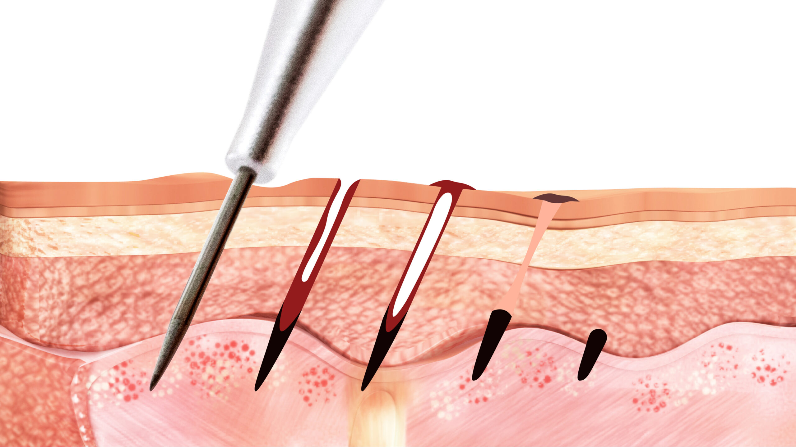
In regenerative wound healing, the skin goes through the following steps:
- First, lymph fluid enters the wound to flush out foreign bodies.
- A fine crust then forms, which comes off after four to six days.
- In the first 24 to 48 hours, the basement membrane closes completely. Any foreign bodies lying under the basement membrane can no longer be pushed out of the wound.
- So-called scavenger cells (macrophage cells) absorb the pigments. As a result, they can no longer pass through the cell walls of the lymphatic system and remain in the tissue together with the pigment.
- If germs have penetrated, the body can render them harmless just a few hours after the injury.
- Hygiene is the top priority during treatment and the healing process.
- The epidermis only regains its original thickness after about four weeks.
Common mistakes during wound healing
Hygiene during and after pigmentation is essential because mistakes in hygiene and care can have a significant impact on the result.
External or mechanical influences on the wound during the healing phase can lead to loss or change of color. The following should therefore be avoided:
- Scratching
- Peeling (directly or in the first few days after pigmentation)
- Scrubbing the pigmented area
- Firm rubbing
- Constant softening of the facial skin with wet compresses
- Excessive washing rituals
- Swimming
- Use of incorrect care products (wound cannot close)
Especially in the first 48 to 72 hours, the artist's aftercare instructions must be followed precisely. The pigmented area must be kept clean, germ-free and dry. Recommend that your clients use hand disinfectant several times a day during the wound healing period (small, pocket-sized hand disinfectants in liquid or gel form are available from any pharmacy or drugstore).
Cornification
The keratinization of the epidermis is individual for each person. The older the skin is, the more horny lamellae there are and the thicker the dead part of the epidermis is. On the palms of the hands and soles of the feet, the horny layer is up to several millimeters thick and is colloquially known as "callus".
The more dead tissue there is, the less the color shines through in pigmented areas of skin. Regular peelings remove the dead horny cells and the color result becomes more radiant again.
Skin color
The natural melanin
The second important factor is the natural melanin or natural skin color. Together with blood circulation (pale to reddish) and carotene (yellow to orange), melanin (light to dark) is decisive for the color tone of the skin.
The number of melanocytes remains constant throughout life, only the amount of melanin produced fluctuates over the years and is dependent on hormones, external influences and sun exposure.
Skin color plays an important role in pigmentation and is decisive in the choice of color. The more melanin there is on a pigmentation, i.e. the darker the epidermis, the more it influences the color intensity of the pigments. The lighter and less keratinized the skin is, the better the brilliance of the color comes into its own. Little sun, care with sun protection factor and gentle peelings protect the result.
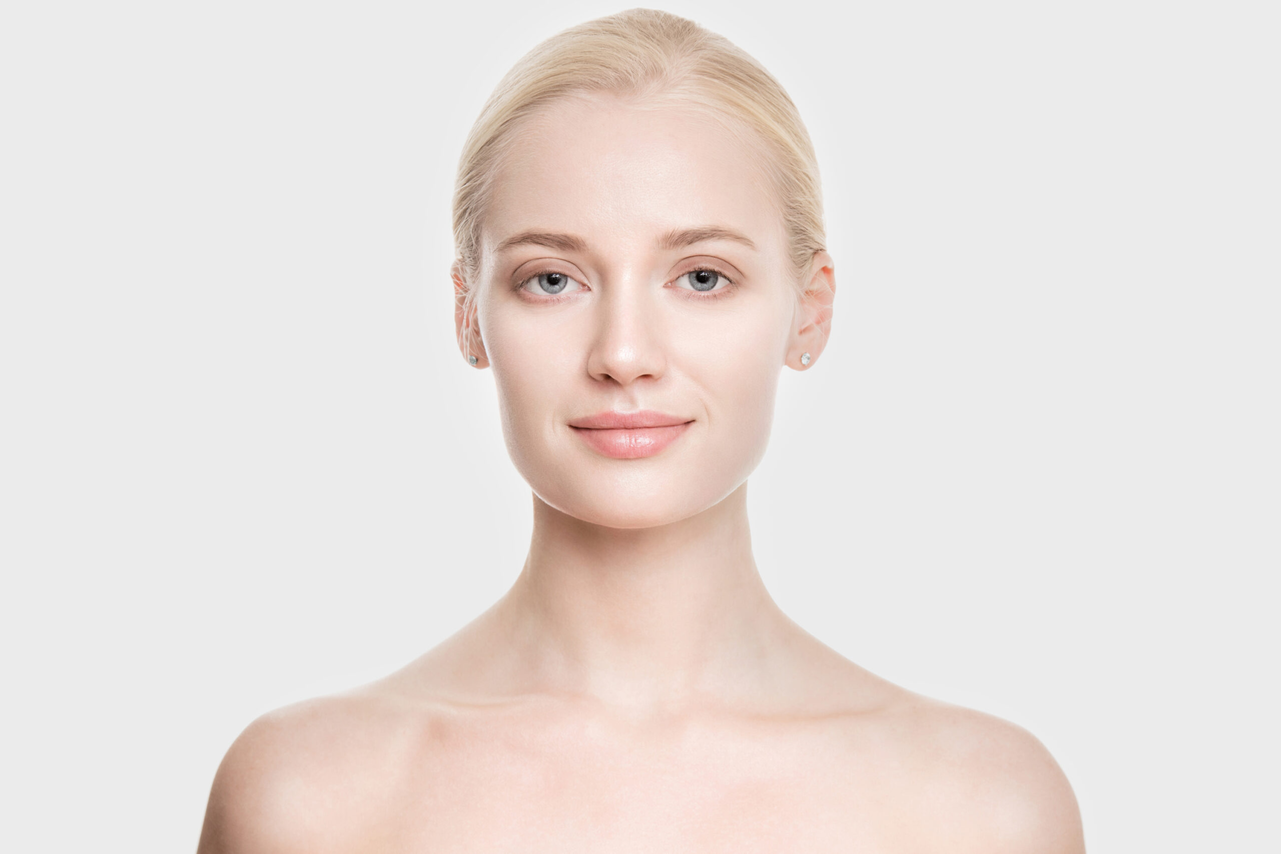
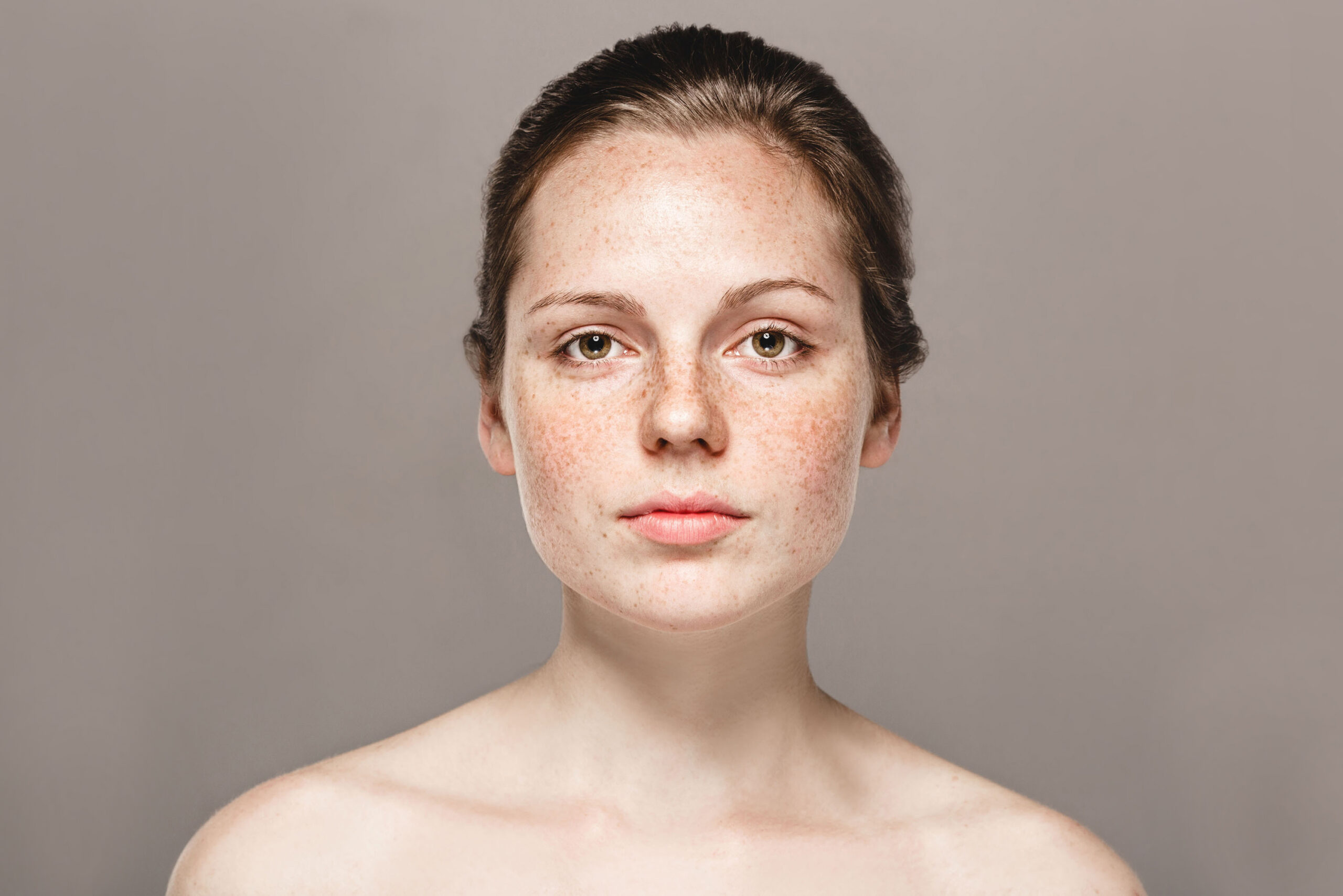
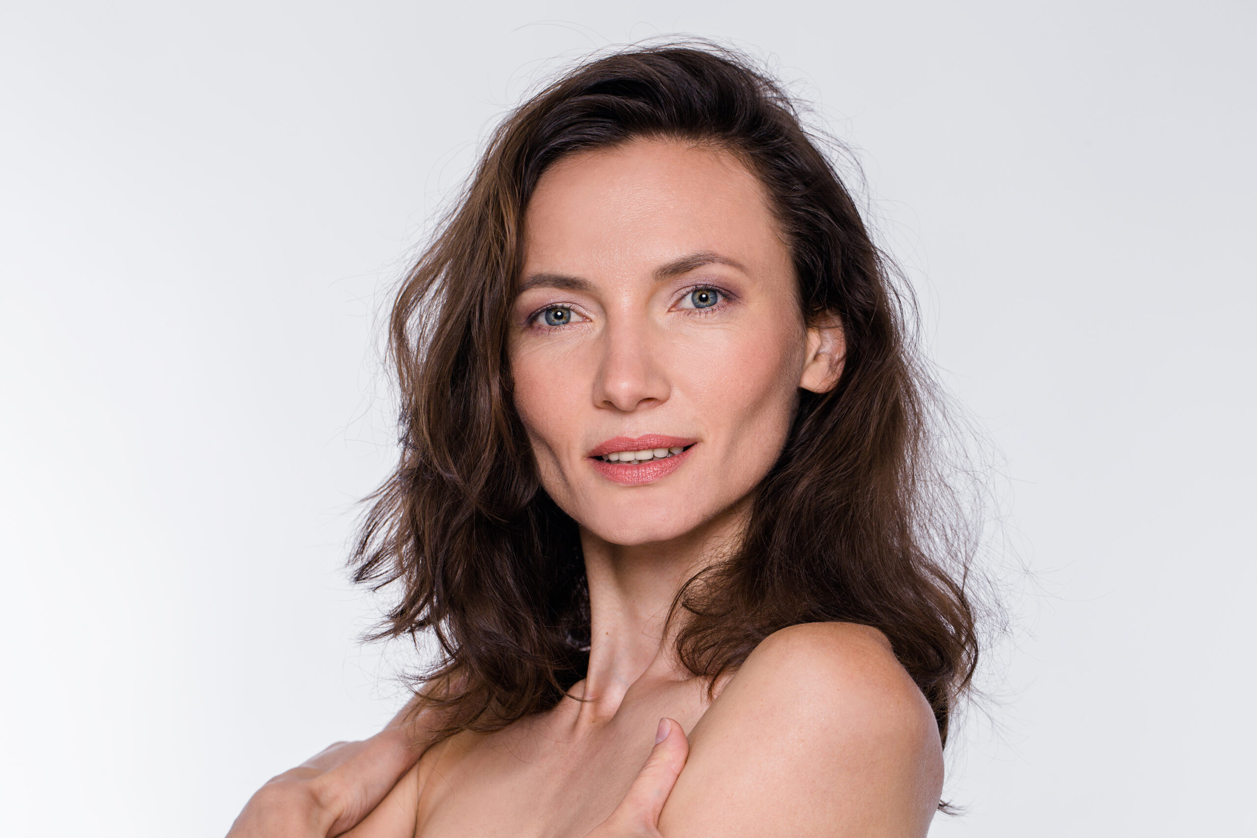
Skin type scale according to Fitzpatrick
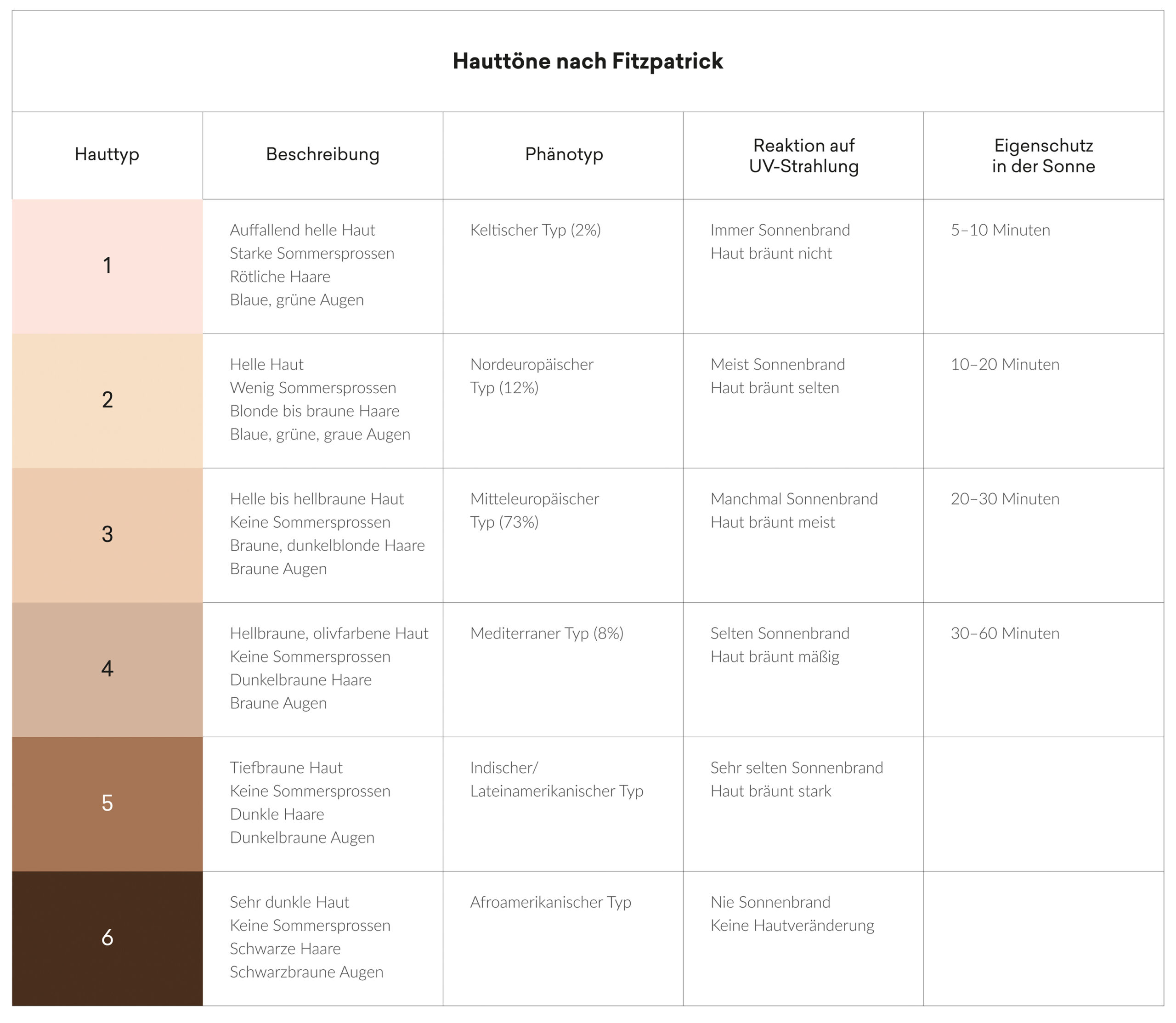
The different skin types are differentiated from light to dark and in undertones such as reddish, olive, yellow, pink, gold, orange, red-orange.
Color direction - undertones
Pink
A pink undertone is found in very light, transparent skin types and is particularly common in Nordic, Irish and Central European regions.
Natural beige
Natural beige is a balanced mixture with no discernible color direction.
Gold/gold-orange
This skin tone appears fresh and healthy due to the slightly tanned skin. This type is often found in South America, southern Europe and Africa.

Red/red-orange
Only a few skin types naturally have this rare undertone. In fair skin types, this undertone is caused by sun exposure, as it makes many tiny dilated veins visible on the skin's surface.
Gold/Olive
These skin types have a high yellow content and little visible blood circulation. This skin type has a sallow appearance and should therefore be pigmented with fresh colors. It is often found in people with Asian, Arabic and Persian roots. As Asian skin contains more collagen than other skin types, it is less prone to wrinkling.

Black Beauty
Special features for dark, oriental, colored and African skin:
Lips
Black African lips look particularly sensual when the contour area, which is usually lighter than the inside of the lips, is only minimally pigmented. The inner area of the lip, on the other hand, is neutralized or lightened with strong and bright orange tones. Depending on the intensity, this is done in one or more treatments.
If there is a desire for intensely coloured lips, the neutralization of the lip color must be completed in order to be able to overpigment with a strong shade. If strong colors are immediately worked into dark skin, they lose their luminosity and brilliance as the dark tissue covers the pigmentation.
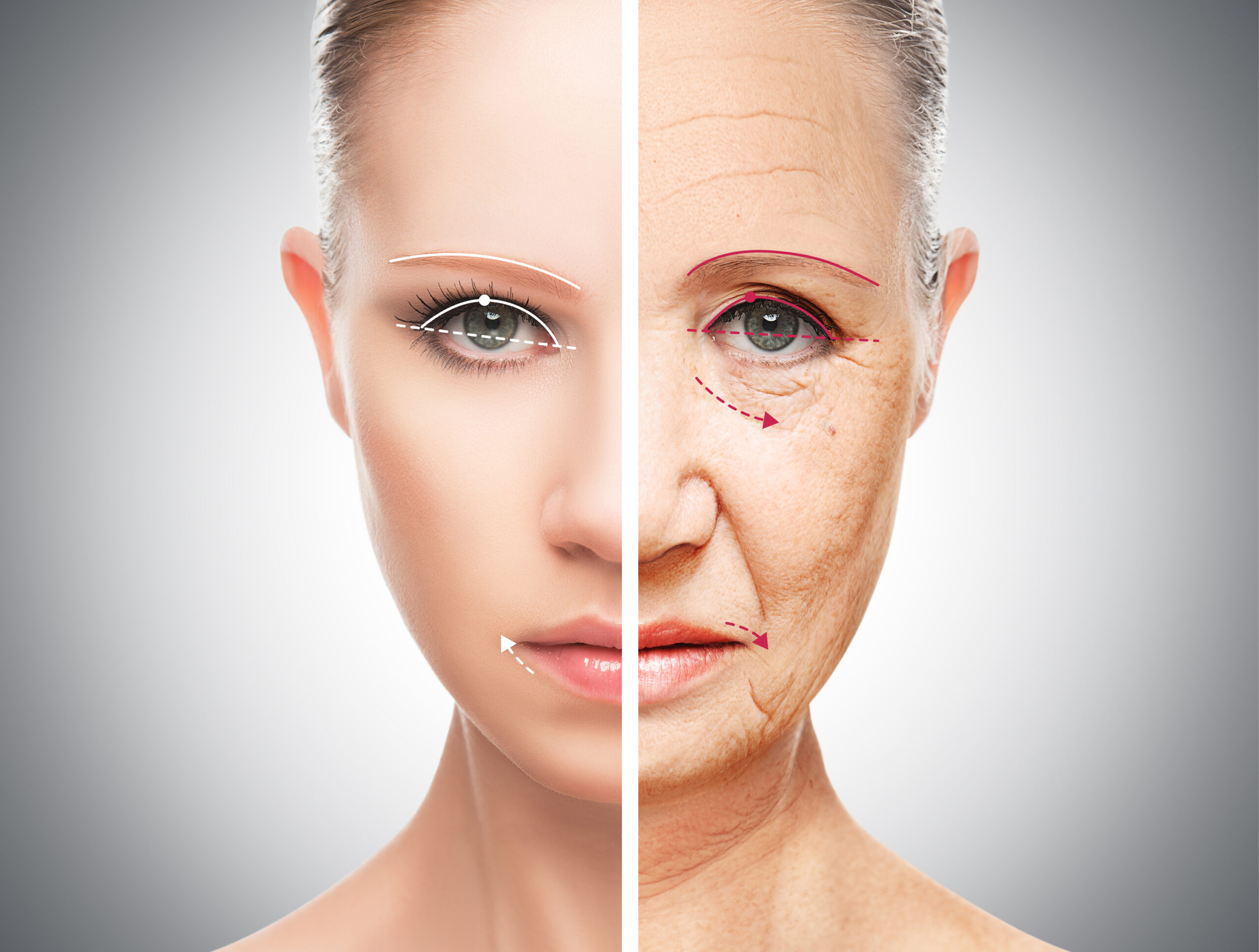
What happens to facial features as we get older?
Practically all customers who opt for permanent make-up as they get older have the same problem: their face changes as their elasticity diminishes. Daily make-up rituals become increasingly difficult and a visual rejuvenation is desired.
The lips
The fullness of the lips decreases slightly over time because less moisture is stored and keratinization increases. This results in a loss of color around the lips, as the keratinized part of the lips no longer allows a view of the blood circulation. They appear frayed, pale and dull.
Copyright © 2017 by Pimp Your Skills GmbH
The copyright for the entire content (text, images and video material) is held by Pimp Your Skills GmbH (unless otherwise stated). The content may not be published, partially published, modified or reproduced without the consent of Pimp Your Skills GmbH.